When a doctor recommends imaging, many patients immediately wonder which scan they actually need. Is an X-ray enough? Is MRI more detailed? Is CT faster? Can ultrasound show the same thing without radiation? These are common questions, especially when symptoms such as pain, swelling, injury, stiffness, abdominal discomfort, or a suspicious finding need a clearer diagnosis.
The simplest answer is that MRI, CT, ultrasound, and X-ray are not better or worse versions of each other. They answer different clinical questions. One test may show bones clearly, another may show soft tissues in more detail, another may be useful for real-time assessment, and another may be preferred when a fast cross-sectional view is needed. The right choice depends on the body area, symptoms, suspected condition, previous findings, urgency, safety considerations, and the type of information the doctor needs.
Through ZagrebMed, patients can send an inquiry for radiology diagnostics such as MRI scan, CT scan, ultrasound, and X-ray imaging. The goal is not to choose a scan randomly, but to connect the symptom or medical question with the most appropriate diagnostic pathway.
When patients start comparing MRI, CT, ultrasound and X-ray
Most people start comparing radiology exams after one of three situations. The first is a new symptom, such as pain after an injury, persistent joint pain, abdominal discomfort, swelling, reduced mobility, or unexplained tenderness. The second is a referral from a doctor who needs imaging before deciding on treatment. The third is a previous finding that needs clarification, follow-up, or a more detailed view.
Patients often ask about imaging when they are dealing with:
- bone pain, suspected fracture, trauma, or joint injury
- back, neck, shoulder, hip, knee, or ankle pain
- tendon pain, muscle injury, or sports-related symptoms
- abdominal, pelvic, thyroid, breast, or soft tissue concerns
- lung, sinus, kidney, urinary, or vascular questions
- preoperative planning or follow-up after treatment
- unclear symptoms where the first examination did not explain the problem
These situations do not automatically mean that the most complex scan is needed. A careful clinical question matters more than the name of the test. For example, X-ray may be the first step after a suspected fracture, ultrasound may be practical for many abdominal or soft tissue concerns, CT may be chosen when speed and detailed cross-sectional anatomy are important, and MRI may be preferred when soft tissue, joints, spine, brain, or complex musculoskeletal structures need detailed evaluation.
Why choosing the right radiology test matters
Radiology is most useful when it answers a specific question. A scan can show anatomy, injury, inflammation, fluid, degeneration, obstruction, bleeding, mass-like changes, or other findings, depending on the method used. But no imaging test sees everything equally well.
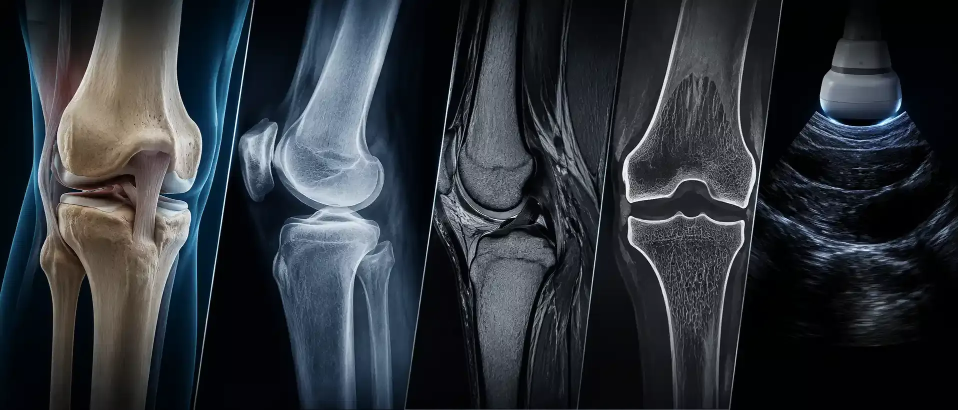
An X-ray is fast and widely used, especially for bones and the chest. CT creates detailed cross-sectional images and can be especially useful when a doctor needs a more complete view of bones, organs, lungs, blood vessels, or acute injury patterns. Ultrasound uses sound waves and is often used for abdominal organs, thyroid, breast, soft tissues, blood flow, and pregnancy-related imaging. MRI uses a magnetic field and radio waves, without ionizing radiation, and is particularly useful for soft tissues, joints, tendons, ligaments, the spine, brain, pelvis, and many complex internal structures.
The right test can shorten the diagnostic pathway. The wrong test can create confusion, require repeat imaging, or fail to answer the question that matters. This is why many imaging decisions are made after a clinical examination, review of previous results, and discussion of symptoms, rather than only based on what the patient feels might be “more detailed.”
What each imaging exam is usually used for
X-ray imaging
X-ray imaging is often used as an initial diagnostic test when the question involves bones, joints, chest structures, certain abdominal findings, or follow-up after injury or treatment. It can help show fractures, bone alignment, joint space changes, some degenerative changes, lung findings, and certain foreign bodies.
X-ray is usually quick and requires little preparation. It does use ionizing radiation, but for many standard X-ray exams the exposure is low. Pregnancy, repeated imaging, and pediatric imaging require extra care and should always be discussed with the referring physician or radiology team.
Ultrasound
Ultrasound uses sound waves to create images in real time. It does not use ionizing radiation and is often chosen for abdominal organs, thyroid, breast, pelvic structures, pregnancy monitoring, soft tissue lumps, fluid collections, and blood flow when Doppler ultrasound is used.
One practical advantage of ultrasound is that the examiner can assess the area dynamically. For some musculoskeletal problems, the patient may be asked to move during the exam so the doctor can observe how a tendon, muscle, or soft tissue structure behaves. Ultrasound is also often used to guide certain needle procedures. Its limitations depend on the body area, patient anatomy, bowel gas, depth of the structure, and the clinical question.
CT scan
A CT scan, also called computed tomography, creates cross-sectional images that can be reconstructed in different planes. CT is often used when doctors need detailed information about bones, lungs, internal organs, trauma, suspected bleeding, kidney stones, certain vascular conditions, or more complex anatomical questions.
CT is usually faster than MRI and can be especially valuable when time matters. Some CT exams use contrast material to show blood vessels, inflammation, tumors, or organ detail more clearly. CT uses ionizing radiation, so the decision should be based on a clear medical indication, especially when repeat scans, pregnancy, or younger patients are involved.
MRI scan
An MRI scan, or magnetic resonance imaging, uses a magnetic field and radio waves to create detailed images. It does not use ionizing radiation. MRI is commonly used for the brain, spine, joints, ligaments, tendons, muscles, pelvis, abdomen, and many soft tissue conditions.
MRI is often selected when the doctor needs to evaluate structures that are not clearly shown on X-ray or when soft tissue detail is central to the diagnosis. It may be useful for persistent joint symptoms, joint stiffness, suspected ligament or meniscus injury, disc problems, nerve compression, soft tissue masses, inflammatory changes, or complex pain that has not been explained by simpler imaging. MRI can take longer than X-ray or CT, and patients with certain implants, devices, metal fragments, claustrophobia, or contrast-related concerns need specific safety screening.
MRI vs CT vs ultrasound vs X-ray: quick practical comparison
A useful way to compare these tests is to think about the question each one is usually asked to answer.
- X-ray: often used for bones, chest imaging, visible fractures, joint alignment, and initial assessment after injury.
- Ultrasound: often used for abdominal organs, thyroid, breast, soft tissues, pregnancy, fluid, and blood flow assessment.
- CT: often used for fast cross-sectional imaging of bones, lungs, organs, trauma, stones, bleeding, and some vascular questions.
- MRI: often used for soft tissues, joints, spine, brain, ligaments, tendons, nerves, pelvis, and detailed musculoskeletal or neurologic evaluation.
The radiation question is also different. X-ray and CT use ionizing radiation. MRI and ultrasound do not use ionizing radiation. This does not automatically make one test “safe” and another “unsafe.” The decision depends on whether the expected diagnostic benefit is greater than the risk, whether another method could answer the same question, and whether contrast material or device-related safety screening is needed.
Preparation also varies. Many X-rays need little preparation. Some ultrasound exams require fasting or a full bladder. CT may require contrast preparation, kidney function checks, or fasting, depending on the exam. MRI may require screening for implants, metal, pacemakers, medication patches, previous surgeries, kidney function if contrast is planned, and comfort issues such as claustrophobia.
What the radiology process usually involves
Phase 1: symptoms and referral question
The process usually starts with the symptom or clinical question. Pain alone is often too broad. A more useful question might be whether there is a fracture, tendon tear, disc herniation, inflammatory change, gallstone, kidney stone, lung finding, mass-like change, or vascular problem. The clearer the question, the easier it is to choose the right imaging method.
Phase 2: choosing the most appropriate test
The doctor or radiologist considers the body area, urgency, previous imaging, age, pregnancy status, implants, kidney function, allergy history, and whether contrast is needed. Sometimes the first test is simple, such as X-ray or ultrasound. In other cases, MRI or CT may be the more appropriate first choice.
Phase 3: preparation before imaging
Before the appointment, patients should bring previous findings, images, discharge letters, medication lists, implant documentation, and referral notes. This is especially useful for MRI safety screening, contrast planning, comparison with earlier scans, and follow-up after surgery or injury.
Phase 4: examination day
During imaging, the radiology team positions the patient and explains what to expect. X-ray and CT are usually faster. Ultrasound depends on the area examined and whether Doppler or dynamic assessment is needed. MRI usually takes longer and requires the patient to remain still while the machine creates image sequences.
Phase 5: report and next step
A radiologist interprets the images and prepares a report. The next step depends on the finding. Some results lead to reassurance and monitoring. Others may require specialist examination, additional imaging, laboratory tests, physical therapy, medication, intervention, or surgical planning. The report should always be interpreted together with symptoms and clinical examination.
Who should discuss imaging choice with a doctor
Patients should avoid choosing imaging only because one test sounds more detailed. A detailed test is not always the right test. A person with a suspected fracture may need X-ray first. A patient with abdominal pain may need ultrasound, CT, laboratory tests, or a different pathway depending on the pattern. A patient with persistent knee pain may need X-ray, MRI, ultrasound, or orthopedic examination depending on age, injury mechanism, swelling, locking, instability, and previous treatment.
Medical discussion is especially important when symptoms are severe, sudden, progressive, or linked with trauma, fever, neurological signs, chest pain, breathing difficulty, unexplained weight loss, cancer history, pregnancy, recent surgery, or significant worsening of function. In urgent situations, patients should seek immediate medical care rather than waiting for a routine imaging appointment.
Imaging is also not always the final answer. Some findings are incidental and may not explain the symptom. Some symptoms are real even if imaging is normal. Some changes seen on imaging are common with age and need clinical interpretation. This is why the radiology report should guide the next step, not replace a medical consultation.
Realistic expectations after imaging
A scan can provide important information, but it cannot always give a complete diagnosis by itself. Imaging results depend on the quality of the clinical question, the body area, the timing of the exam, technical factors, and how the finding matches the patient’s symptoms.
Patients should expect one of several possible outcomes. The scan may confirm a suspected diagnosis. It may rule out an important problem. It may show a finding that needs follow-up. It may suggest a different test. It may show age-related or old changes that need correlation with symptoms. In some cases, a doctor may recommend conservative treatment first and imaging only if symptoms persist or worsen.
A practical preparation checklist can help before any radiology appointment:
- Bring previous imaging reports and images if available.
- Write down the main symptom, when it started, and what makes it better or worse.
- Tell the team about pregnancy, implants, pacemakers, metal fragments, allergies, kidney disease, or previous contrast reactions.
- Ask whether fasting, a full bladder, medication changes, or blood tests are needed before the exam.
- Confirm how and when the radiology report will be available.
Next step through ZagrebMed
If you are unsure which radiology exam fits your symptoms or previous findings, the most useful next step is to send an inquiry with your medical question, previous reports, and the body area that needs assessment. ZagrebMed can help connect the inquiry with available radiology services and relevant partner clinics in Zagreb.
Depending on the exam and indication, radiology diagnostics are available through ZagrebMed partners such as Agram, Sinteza, and Akromion. The goal is to help the patient move from uncertainty to the right diagnostic step, without unnecessary waiting and without choosing a test without clinical context.
This content is informational and does not replace a medical consultation, diagnosis, or individualized treatment recommendation. A doctor or radiologist should decide which imaging test is appropriate based on your symptoms, medical history, examination, and previous findings.
Sources
- RadiologyInfo.org, American College of Radiology and Radiological Society of North America: Magnetic Resonance Imaging (MRI)
- RadiologyInfo.org, American College of Radiology and Radiological Society of North America: Computed Tomography
- RadiologyInfo.org, American College of Radiology and Radiological Society of North America: Ultrasound
- RadiologyInfo.org, American College of Radiology and Radiological Society of North America: X-ray Radiography
- American College of Radiology: ACR Appropriateness Criteria
- U.S. Food and Drug Administration: Medical X-ray Imaging